
Page 14 - Dentity_EN_0910_high
P. 14
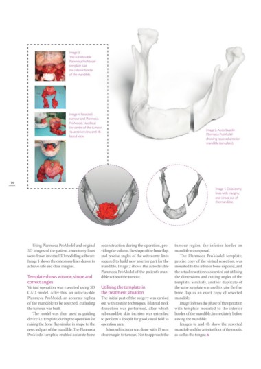
Image 3. Image 2. Autoclavable
The autoclavable Planmeca ProModel
Planmeca ProModel showing resected anterior
template is at mandible (template).
the inferior border
of the mandible. Image 1. Osteotomy
lines with margins,
Image 4. Resected and virtual cut of
tumour and Planmeca the mandible.
ProModel. Needle at
the centre of the tumour.
4a. anterior view, and 4b
lateral view.
14
Using Planmeca ProModel and original reconstruction during the operation, pro tumour region, the inferior border on
3D images of the patient, osteotomy lines viding the volume, the shape of the bone flap, mandible was exposed.
were drawn in virtual 3D modelling software. and precise angles of the osteotomy lines
Image 1 shows the osteotomy lines drawn to required to build new anterior part for the The Planmeca ProModel template,
achieve safe and clear margins. mandible. Image 2 shows the autoclavable precise copy of the virtual resection, was
Planmeca ProModel of the patient’s man‑ mounted to the inferior bone exposed, and
Template shows volume, shape and dible without the tumour. the actual resection was carried out utilising
correct angles the dimensions and cutting angles of the
Utilising the template in template. Similarly, another duplicate of
Virtual operation was executed using 3D the treatment situation the same template was used to raise the free
CAD model. After this, an autoclavable bone flap as an exact copy of resected
Planmeca ProModel, an accurate replica The initial part of the surgery was carried mand ible.
of the mandible to be resected, excluding out with routine techniques. Bilateral neck
the tumour, was built. dissection was performed, after which Image 3 shows the phase of the operation
submandible skin incision was extended with template mounted to the inferior
The model was then used as guiding to perform a lip split for good visual field to border of the mandible, immediately before
device, i.e. template, during the operation for operation area. sawing the mandible.
raising the bone flap similar in shape to the
resected part of the mandible. The Planmeca Mucosal incision was done with 15 mm Images 4a and 4b show the resected
ProModel template enabled accurate bone clear margin to tumour. Not to approach the mandible and the anterior floor of the mouth,
as well as the tongue.