
Page 11 - Dentity_EN_0910_high
P. 11
11
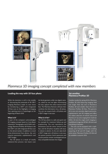
Planmeca 3D imaging concept completed with new members
Leading the way with 3D face photo Yet another
Planmeca ProMax 3D
While the dentistry is still in the middle the facial geometry while two digital came- Another novelty for spring 2010 is Planmeca
of discovering the potentials of 3D CBVT ras, aided by two led lights illuminating ProMax 3D Mid featuring imaging field
imaging Planmeca made its next move the area, capture the colour texture of the size larger than the one in Planmeca
by introducing a CBVT unit integrated face. The Planmeca Romexis software then ProMax 3D and smaller than the one in
3D face scan at the Highlights in Imaging combines the information into a 3D photo. Planmeca ProMax 3D Max. This genuine
Tour opening event in Copenhagen at the The unit can acquire both a 3D photo and all-in-one unit provides digital panoramic,
beginning of March 2010. a CBVT image in one scan. cep halometric and 3D imaging and offers
the widest selection of volume sizes of all
What is it? What is it for? Planmeca X-ray units ranging from small size
(Ø40 x 50 mm) for single tooth imaging to
As part of the company’s acknowledged The 3D face photo is a safe and quick tool maxillofacial image size (Ø160 x 90 mm) and
3D imaging concept based on Cone Beam for example for treatment follow-up and everything in between.
Volumetric Tomography and SCARA tech- preplanning. The unit reproduces the
nology, Planmeca ProMax 3D ProFace is the dimensions and the colour texture of the The unit can be equipped with a cep h
first CBVT unit with integrated 3D face scan face, and the 3D photo visualises soft tissue alostat, and it features SmartPan enabling
on the dental market. In addition to novel in relation to dentin. As the unit takes both acquiring of 3D and 2D images with the
three-dimensional face photo, the unit a CBVT image and a 3D photo in one scan, same sensor. Planmeca ProMax 3D Mid will
acquires panoramic, cephalometric and the patient position, facial expression and be available in autumn 2010.
CBVT images. muscle position remain unchanged and are
thus compatible between the images.
The 3D photo is a result of totally
radiation-free process: two lasers scan