
Page 10 - Dentity_EN_0910_high
P. 10
3D cone beam imaging Conclusions
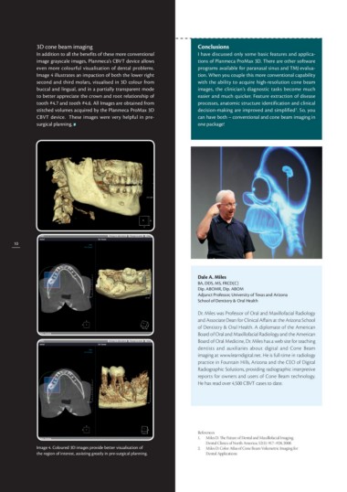
In addition to all the benefits of these more conventional I have discussed only some basic features and applica-
image grayscale images, Planmeca’s CBVT device allows tions of Planmeca ProMax 3D. There are other software
even more colourful visualisation of dental problems. programs available for paranasal sinus and TMJ evalua-
Image 4 illustrates an impaction of both the lower right tion. When you couple this more conventional capability
second and third molars, visualised in 3D colour from with the ability to acquire high-resolution cone beam
buccal and lingual, and in a partially transparent mode images, the clinician’s diagnostic tasks become much
to better appreciate the crown and root relationship of easier and much quicker. Feature extraction of disease
tooth #4.7 and tooth #4.6. All Images are obtained from processes, anatomic structure identification and clinical
stitched volumes acquired by the Planmeca ProMax 3D decision-making are improved and simplified2. So, you
CBVT device. These images were very helpful in pre- can have both – conventional and cone beam imaging in
surgical planning. one package!
10 Dale A. Miles
Image 4. Coloured 3D images provide better visualisation of BA, DDS, MS, FRCD(C)
the region of interest, assisting greatly in pre-surgical planning. Dip. ABOMR, Dip. ABOM
Adjunct Professor, University of Texas and Arizona
School of Dentistry & Oral Health
Dr. Miles was Professor of Oral and Maxillofacial Radiology
and Associate Dean for Clinical Affairs at the Arizona School
of Dentistry & Oral Health. A diplomate of the American
Board of Oral and Maxillofacial Radiology and the American
Board of Oral Medicine, Dr. Miles has a web site for teaching
dentists and auxiliaries about digital and Cone Beam
imaging at www.learndigital.net. He is full-time in radiology
practice in Fountain Hills, Arizona and the CEO of Digital
Radiographic Solutions, providing radiographic interpretive
reports for owners and users of Cone Beam technology.
He has read over 4,500 CBVT cases to date.
References
1. Miles D. The Future of Dental and Maxillofacial Imaging.
Dental Clinics of North America; 52(4): 917–928, 2008.
2. M iles D: Color Atlas of Cone Beam Volumetric Imaging for
Dental Applications