
Page 8 - Dentity_EN_0910_high
P. 8
What are the diagnostic tasks that CBVI simplifies?
What are those CBVI will replace?
In December 2008, I published an article a line pair resolution of 7 lp/mm the clinician
in the Dental Clinics of North America is likely to require much fewer intraoral With Planmeca ProMax 3D
that answers questions I receive frequently images. And, as the examples on Images 3a you also have:
from dentists trying to understand CBVI and 3b demonstrate, these “super bitewings”
technology and its applications1. I made also give the dentist very good periapical 1. c onventional panoramic program
some predictions based on answers to their views of all teeth from the canine to third
2. i mproved interproximal
questions. molar. This is very exciting!
Pan program
In my opinion, CBVI will be the “standard Imagine avoiding intraoral images on
of care” for those diagnostic tasks listed in a 3-year-old. They don’t like anything in their 3. i mproved orthogonal
Table 1. The additional applications for mouth at that age. You can see what you Pan program (periodontal)
which CBVI can be used are in Table 2. From need to see, including tooth development on
these tables you will see that CBVI has 2 low dose, magnified super bitewings! 4. bitewing program mode
already impacted every aspect of dental (interproximality).
As one pediatric dentist colleague stated,
imaging. Machines that are multi-functional, “I may never have to place an X-ray receptor
such in a child’s mouth again”. His reasoning was
as Planmeca ProMax 3D, appear to be the that the image quality was so good that he
devices that best match the current and could see all the diagnostic features he
future needs of the dental profession. needed. The primary enamel is so thin, that
When you examine the tables, you see a cavity on a primary tooth is a cavity! He’s
that there are still some tasks that cone beam not looking for the very early lesions most
imaging will not replace, for instance, inter- times in the very thin outer enamel. He’s
proximal carious lesion detection. Despite evaluating the furcation areas, tooth devel-
the low dose and small voxel size, caries opment and anomalies which might
detection will still require conventional be present. Take a look for yourself at
intraoral images like bitewings. However the excellent image quality you get with
8 with Planmeca ProMax 3D’s capability to do a super bitewing.
over-sized digital bitewing projections at
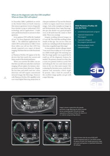
Image 2a shows a typical thin slice pseudo-
panoramic image depicting the inferior alveolar
nerves contacting teeth #3.8 and 4.8. Image
captured on a Planmeca ProMax 3D: three 8 x 8
cm volumes using stitching software.
Image 2b shows that the nerve DOES NOT
contact impacted tooth #4.7, but DOES contact
the apices of tooth #4.8. Pseudo-panoramic image
and canal marking done in CyberMed OnDemand
3D software.