
Page 7 - PlanWorld_1_2013_high
P. 7
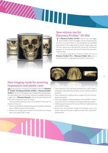
New volume size for
                                                                           Planmeca ProMax® 3D Mid

                                                                           The Planmeca ProMax® 3D Mid X-ray unit has a new, bigger
                                                                               sensor, thanks to which the biggest volume size generated by
                                                                           the unit is now up to 20x18 cm. This also signifies a considerable
                                                                           improvement in the image quality as well as a larger target area.
                                                                           The new volume size is well suited for instance for orthodontic
                                                                           purposes. At the same time, the size of the SmartPan panoramic
                                                                           image increases.

                                                                              A new sensor generation will also be taken into use in

                                                                           .Planmeca ProMax® 3D and Planmeca ProMax® 3D s units, so

                                                                           the improvement in image quality also applies to these units

                                                                                                                                                  7

New imaging mode for scanning                                              •	 Utilise impression scans and those combined with a CBCT image in
impressions and plaster casts                                                orthognatic surgery planning and follow-up (pre- and post-operative
                                                                             situations).
Planmeca is proud to introduce a new imaging mode to the Planmeca
    ProMax® 3D, Planmeca ProMax® 3D Mid and Planmeca ProMax®               •	 Create snapshots for making deviation images of the pre- and post-
3D Max X-ray units. The imaging mode is designed for scanning impres-        operative situations.
sions and plaster casts. The scanned impression is available instantly as
a digital cast in Planmeca Romexis® and can be superimposed with           •	 Export the mapped or separate impression scans and CBCT volumes
CBCT data or exported in open STL format.                                    in open STL format to orthodontic and surgical planning software.

   The acquired 3D impression scans can be utilised in many ways. For      •	 Utilise impression scans and 3D crowns in Romexis implant planning
example, the user can:                                                       with the CBCT volume.
•	 Follow the orthodontic treatment progress by comparing impressions
                                                                           •	 Benefit from the automatic storage of impression casts in the
  taken in different phases of the treatment.                                patient’s data with all other images (intraoral, CBCT, panoramic,
•	 Measure the changes and movements of the teeth and compare                cephalometric).

  them to the original CBCT image.