
Page 14 - PlanWorld_1_2012_high
P. 14
New potential with mobile,
low-dose 3D imaging
The careful development process proved
worth the effort as Planmed Verity turned
out as the first ever mobile 3D extremity
imaging device, which currently has no real
competitors. According to Seppälä, when
a patient nowadays comes to a clinic with
an injured extremity, the standard procedure example, in a 2D image of the forefoot,
is to take a 2D. In many cases, however, the numerous small bones show as
the injury does not show in a 2D image or overlapping, making it difficult to see how
a lot is left to guesswork. a bone is bent or how a single articular
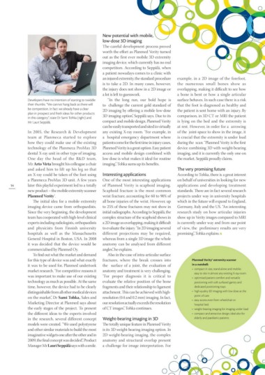
Developers have no intention of starting to twiddle “In the long run, our bold hope is surface behaves. In such case there is a risk
their thumbs. “We cannot hang back as there will to challenge the current gold standard of that the foot is diagnosed as healthy and
be competition. In fact we already have a clear 2D imaging by offering a mobile low-dose the patient is sent home with an injury. By
plan in prospect and fresh ideas for other products 3D imaging option”, Seppälä says. Due to its comparison, in 3D CT or MRI the patient
in this category”, state Dr Sami Tohka (right) and compact and mobile design, Planmed Verity is lying on the bed and the extremity is
Mr Lauri Seppälä. can be easily transported and sited in virtually at rest. However, in order for a arrowing
In 2005, the Research & Development any existing X-ray room. “For example, in of the joint-space to show in the image, it
team at Planmeca started to explore a hospital emergency department where is crucial that the extremity is under load
how they could make use of the existing patients come for the first time in injury cases, during the scan. “Planmed Verity is the first
technology of the Planmeca ProMax 3D Planmed Verity is a great option. Easy patient device combining 3D with weight-bearing
dental X-ray unit in other type of imaging. access and mobile design combined with imaging, and it is currently the only one on
One day the head of the R&D team, low dose is what makes it ideal for routine the market, Seppälä proudly claims.
Mr Arto Virta brought his colleague a chair imaging”, Tohka sums up its benefits. The very promising future
and asked him to lift up his leg so that
an X-ray could be taken of the foot using Interesting applications According to Tohka, there is a great interest
a Planmeca ProMax 3D unit. A few years One of the most interesting applications on behalf of universities in looking for new
14 later this playful experiment led to a totally of Planmed Verity is scaphoid imaging. applications and developing treatment
new product – the mobile extremity scanner Scaphoid fracture is the most common standards. There are in fact several research
Planmed Verity®. wrist fracture, accounting for 60 to 90% of projects under way in university hospitals
The initial idea for a mobile extremity all bone injuries of the wrist. However, up which in the future will expand to England,
imaging device came from orthopaedists. to 25% of these fractures may not show in Germany, Italy and the US. “An interesting
Since the very beginning, the development initial radiographs. According to Seppälä, the research study on how articular injuries
team has cooperated with high-level clinical complex structure of the scaphoid shows in show up in Verity images compared to MRI
experts including radiologists, orthopaedists a 2D image as overlapping, making it difficult is currently under way and from our point
and physicists from Finnish university to evaluate the injury. “In 2D imaging several of view, the preliminary results are very
hospitals as well as the Massachusetts different projections may be required, promising”, Tohka explains.
General Hospital in Boston, USA. In 2008 whereas from a single 3D image the whole
it was decided that the device would be anatomy can be analysed from different
commercialised by Planmed Oy. angles”, he explains.
To find out what the market and demand Also in the case of intra-articular surface
for this type of device was and what exactly fractures, where the break crosses into Planmed Verity® extremity scanner
it was to be used for, Planmed undertook the surface of a joint, the evaluation of in a nutshell:
market research. “For competitive reasons it anatomy and treatment is very challenging. • compact in size, stand-alone and mobile:
was important to make use of our existing “For proper diagnosis it is critical to easy to site in almost any existing X-ray room
technology as much as possible. At the same evaluate the relative position of the bone • optimised patient comfort and versatile
positioning with soft surfaced gantry and
time, however, the device had to be clearly fragments and their relationship to ligament dedicated positioning trays
distinguishable from all other medical devices attachment. This can be achieved with high- • high-quality 3D imaging with low dose at the
on the market”, Dr Sami Tohka, Sales and resolution (0.4 and 0.2 mm) imaging. In fact,
Marketing Director at Planmed says about our resolution actually exceeds the resolution point of care
the early stages of the project. To present of CT images”, Tohka continues. • easy access even from wheelchair or
hospital bed
• weight-bearing imaging for imaging under load
the different ideas to the experts involved • compact and attractive design; ideal also for
elderly and paediatric patients
in the research, several different concept Weight-bearing imaging in 3D
models were created. “We used polystyrene The totally unique feature in Planmed Verity
and other similar materials to build the most is its 3D weight-bearing imaging option. In
imaginative widgets one after the other and in 2D weight-bearing imaging, the complex
2009, the final concept was decided”, Product anatomy and structural overlap present
Manager Mr Lauri Seppälä says with a smile. a challenge for image interpretation. For